

What Is The Function Of The Cytosol In An Animal Cell
What is a cell membrane? A quick overview
Our cell is literally a balloon filled with water (70% of our body weight is h2o). This soft just tough balloon is made from the cell membrane (also known as the plasma membrane). The cell membrane is a thin biological membrane that separates the interior of cells from the exterior space and protects the cells from the surrounding environment.
The cell membrane is made of two layers of lipid films (oil molecules) with many kinds of proteins inserted. These proteins control the movement of molecules such as water, ions, nutrients, and oxygen in and out of the cell.
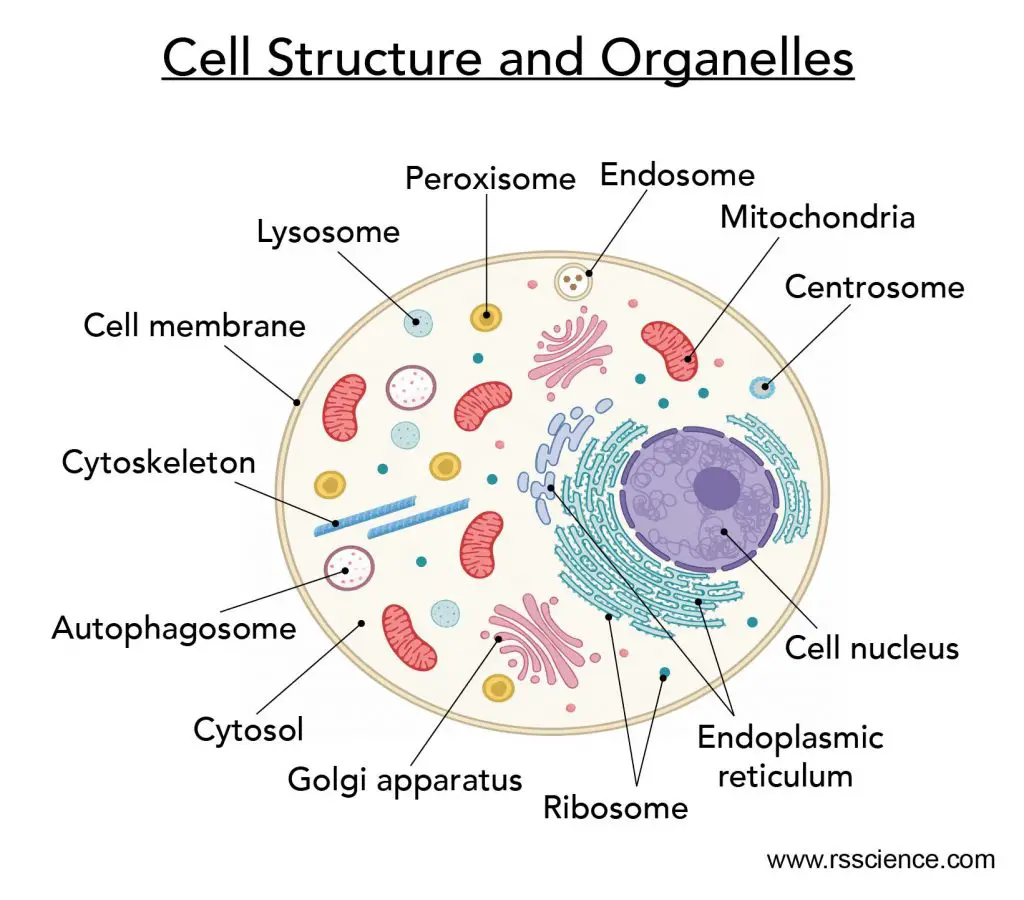
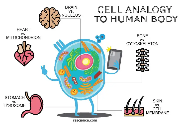
[In this effigy] The anatomy of an animal jail cell with organelles labeled.
Enclosed by this cell membrane are the cell'south constituents, including cell organelles and jelly-like fluids chosen cytosols with water-soluble molecules such as proteins, nucleic acids, carbohydrates, and substances involved in cellular activities.
The prison cell membrane, therefore, has two key functions:
i. To exist a barrier keeping the constituents of the cells in and unwanted substances/toxics out.
2. To exist a gate allowing the transportation of essential nutrients into the cells and waste products out of the cells.
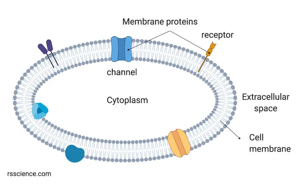
[In this figure]The prison cell membrane defines the within and outside spaces of a cell.
The cell membrane is a phospholipid bilayer membrane with many proteins. These proteins could be inserted in or associated with the membrane and function every bit channels (decision-making in and out of molecules) or receptors (receiving signals from the outside world).
The prototype was created with BioRender.com.
The structure of cell membrane
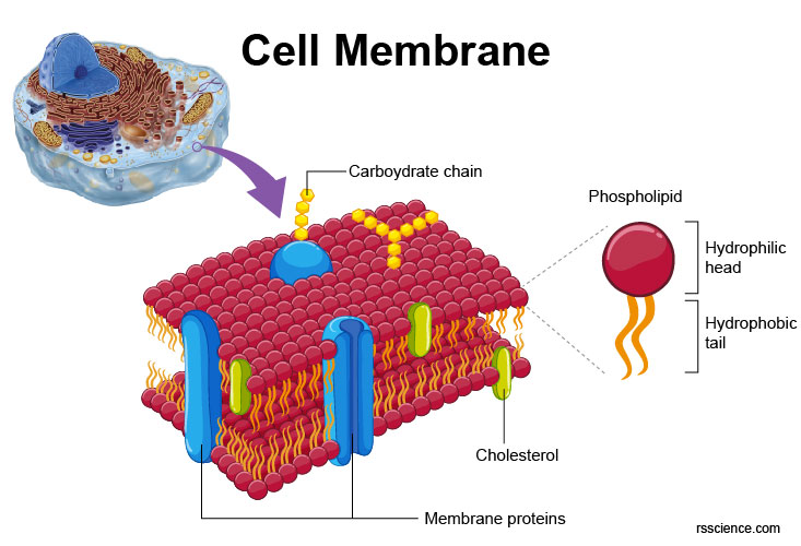
[In this figure] The fluid mosaic model of the cell membrane showing membrane proteins assembled with a lipid bilayer.
Phospholipid bilayer equally a versatile biological bulwark
The backbone structure of the cell membrane is a thin polar membrane made of two layers of lipid molecules, called lipid bilayer (or phospholipid bilayer). This bilayer is formed past the amphiphilic phospholipids, which have a hydrophilic (preferring water) phosphate head and a hydrophobic (preferring to stay away from water) tail consisting of two fatty acid chains.
In an aqua environment, the hydrophobic tails of many phospholipids naturally stay together with their hydrophilic phosphate heads facing the exterior water molecules. The lipid bilayer forms spontaneously by self-assembly.
Some other blazon of lipid molecule, called steroid, is as well a central function of the cell membrane. Cholesterol is the near common steroid and the level of cholesterol tin can potentially alter the fluidity and function of the membrane.
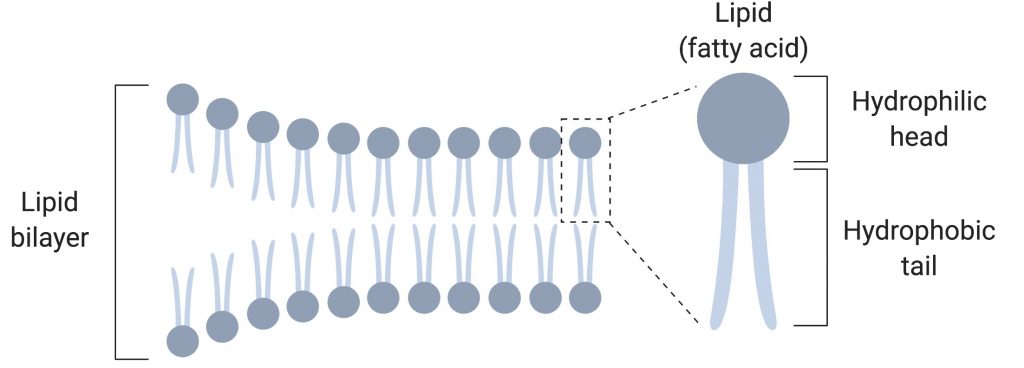
[In this figure]The cell membrane is made of ii lipid films, called lipid bilayer.
Selective permeability of cell membrane
The lipid bilayers are ideally suited to keeps ions, proteins, and other charged molecules from diffusing across the membrane, even though they are only a few nanometers in width. At the aforementioned time, uncharged molecules and gases tin can easily cantankerous the cell membrane. This property is chosen semi-permeability or selective permeability.
Selective permeability prevents gratis diffusion of molecules so that membranes can class compartments that keep distinct internal and external environments. This is because that the hydrophobic cores of lipid bilayers (created past the fatty acid chains) are impermeable to nearly water-soluble (hydrophilic) molecules. Withal, small molecules without electric charges, such every bit COii, Due north2, Otwo, and molecules with high solubility in fatty such every bit ethanol, tin can cross membranes about freely.
This belongings allows cells to regulate common salt concentrations and pH inside the cells. Transportation of ions across their membranes requires special permission by proteins called ion pumps or channels.
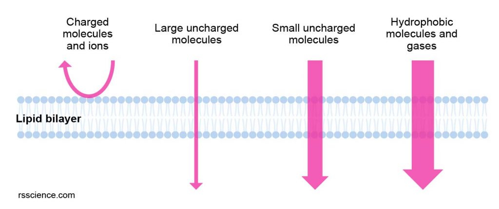
[In this effigy]Selective permeability of jail cell membrane: the size and electric features of molecules determine their ability to cross jail cell membranes.
Pocket-sized molecules without electrical charges (e.yard., gases) and oil-soluble molecules can cross membranes virtually freely. Permeability is lower for uncharged molecules such as h2o and glycerol. The ability of large uncharged molecules, such equally glucose, to cross membranes is low. Membranes are highly impermeable to ions and charged molecules.
Phospholipid bilayers are widely used for all membrane-leap organelles
Since the lipid bilayers are so neat to create compartments for biochemical reactions, the membrane-leap organelles (such as the nucleus, endoplasmic reticulum, mitochondria, chloroplasts, Golgi apparatus, lysosomes, peroxisomes, and vacuoles) all use the aforementioned lipid bilayers for their membranes. We even create human being-fabricated spherical-shaped vesicle that is composed of lipid bilayer membranes, called liposomes, to assist united states of america evangelize drugs and vaccines.

[In this effigy] The iii principal structures phospholipids form in solution: the liposome (a airtight bilayer), the micelle, and the bilayer.
Prototype credit: wiki
Membrane proteins
In that location are various kinds of proteins associated with the cell membrane to perform many essential biological functions. These proteins can contribute to around 50% of membrane volume. Approximately a 3rd of the genes in multicellular organisms code specifically for membrane-related proteins.
Membrane proteins consist of 3 main types: transmembrane proteins, lipid-anchored proteins, and peripheral proteins.
| Type | Description | Examples |
| Transmembrane proteins | These proteins are inserted in the lipid bilayers with one or two parts facing either extracellular or intracellular spaces. | Ion channels, Chiliad protein-coupled receptors |
| Lipid anchored proteins | The protein itself is non in contact with the membrane. Instead, they ballast on the cell membrane through covalently bound to lipid molecules. | Chiliad proteins |
| Peripheral proteins | Fastened to integral membrane proteins or associated with peripheral regions of the lipid bilayer. These proteins tend to have merely temporary interactions with cell membranes for certain reactions. | Some enzymes and hormones |
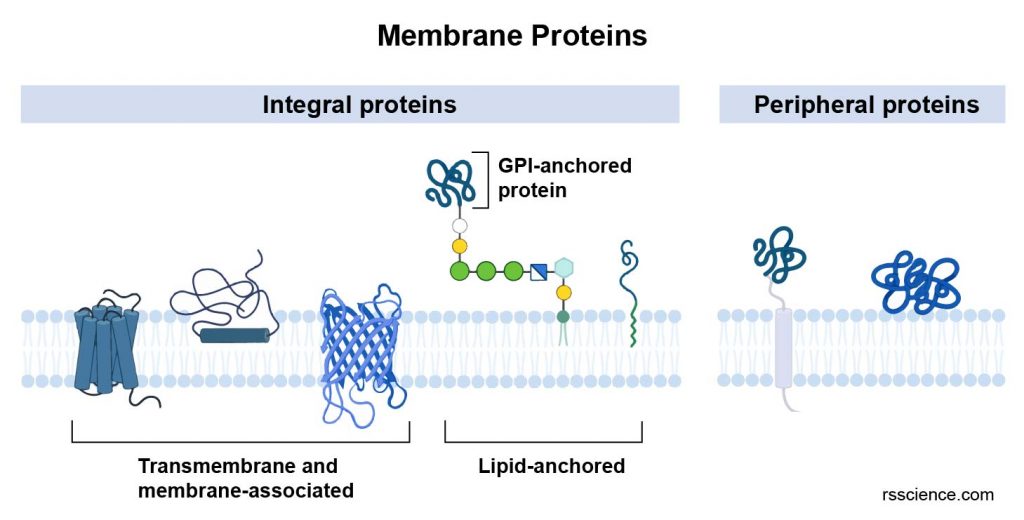
[In this effigy] Schematic diagram of transmembrane, peripheral, and lipid-anchored membrane proteins.
Some transmembrane proteins present carbohydrate bondage on the cell's outer surface (as well called glycoproteins).
Membrane proteins enable the cell membranes to exercise diverse functions. Nosotros volition hash out these functions after.
Fluid mosaic model: What makes the cell membrane fluid?
The jail cell membrane is flexible and fluidic. To better describe the properties of the cell membrane, scientists explicate the jail cell membrane appearance and functions using the fluid mosaic model.
If yous zoom in on the cell membrane, you will see the bounding main of lipid molecules decorated with membrane proteins, cholesterols, and carbohydrates. These molecules are constantly moving in ii dimensions, in a fluid fashion, like to icebergs floating in the ocean. There is no consistent blueprint or organization of these molecules; they are more like a mosaic.
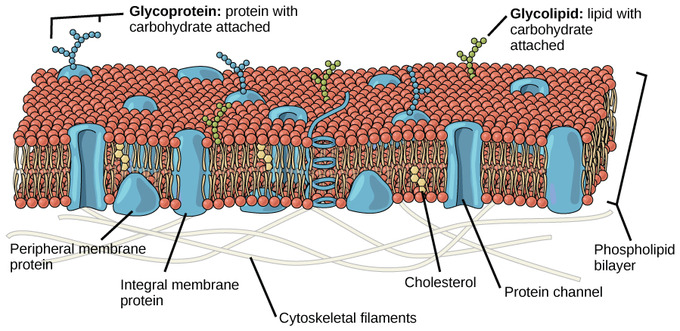
[In this figure] The fluid mosaic model of the cell membrane describes the jail cell membrane as a fluid combination of phospholipids, cholesterol, and proteins.
Photo source: Biology LibreTexts
The fluid property of the cell membrane is essential for many activities of cells. At that place are iii main factors that influence cell membrane fluidity:
(1) Temperature: The temperature can affect how the phospholipids movement and how close they stay. When it's cold, they are closer together and when information technology's hot, they move farther apart.
(2) Cholesterol: The cholesterol molecules are randomly distributed in the lipid bilayer, helping the membrane stay fluid. The cholesterol holds the phospholipids together at a proper distance, not too close and not too far. Without cholesterol, the phospholipids start to separate from each other, leaving large gaps at a warm temperature. On the other hand, without cholesterol, the phospholipids in your cells will showtime to get closer together when exposed to common cold, making it more hard for small molecules, such as gases, to squeeze through the phospholipids similar they normally do.
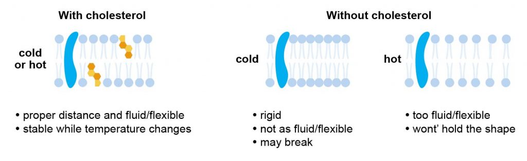
[In this effigy] Cholesterol is a cardinal component of the cell membrane. The presence of cholesterol molecules can stabilize the properties of prison cell membrane across a range of temperatures.
(3) Saturated and unsaturated fatty acids: Fatty acids are phospholipid tails. Saturated fat acids are bondage of carbon atoms that have only unmarried bonds betwixt them. Equally a result, the chains are straight and easy to pack tightly.
Unsaturated fatty acids are bondage of carbon atoms that have double bonds between some of the carbons. The double bonds create kinks inside the bondage, making it harder for the bondage to pack tightly. These kinks play a part in membrane fluidity because they increase the space betwixt the phospholipids, making the molecules harder to freeze at lower temperatures. In addition, the increased space allows certain small molecules, such equally CO2 to cross the membrane chop-chop and easily.
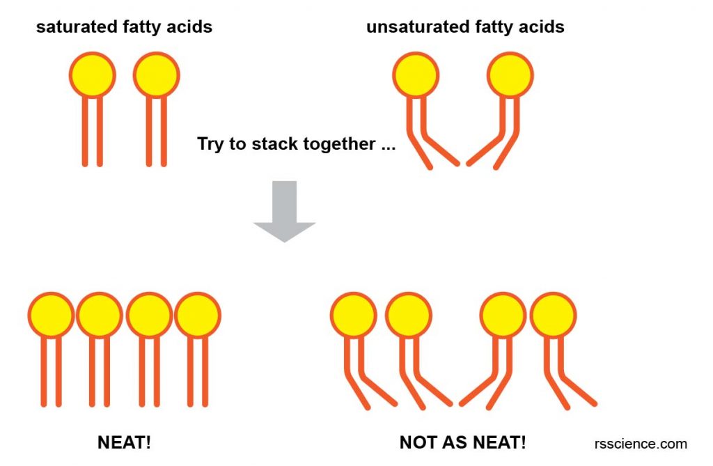
[In this effigy] The limerick of saturated and unsaturated fatty acids affects the fluidic of the prison cell membrane.
What does the cell membrane do? – The jail cell membrane function
Jail cell membrane serves as a barrier of cells, just like our skin
The jail cell membrane encloses the cytoplasm of living cells, physically separating the intracellular components from the extracellular environment. Establish cells possess prison cell walls exterior the cell membrane for actress protection and back up.
Cell membrane defines the cell shape and provides the sites for cell anchorage
The cell membrane plays a office in anchoring the cytoskeleton to provide shape to the cell (particularly in beast cells without the cell wall). The jail cell membrane also provides the sites to interact with the extracellular matrix and other cells. These contact points can transmit the extracellular mechanical stimuli (similar pressure or shear strength) into the cytoskeleton, resulting in the change of cell behavior by altering gene expression in the nucleus.
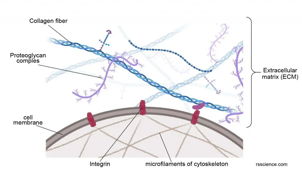
[In this figure] The cell membrane and transmembrane proteins serve as attachment points to bring intracellular cytoskeleton and extracellular matrix (ECM) together.
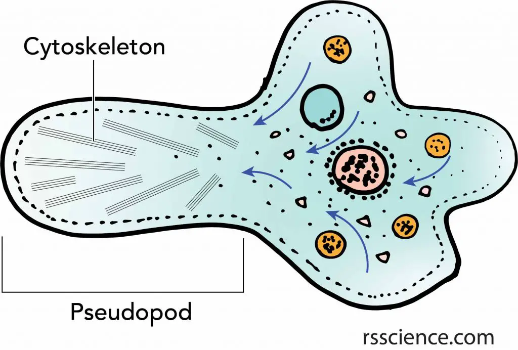
[In this figure] Amoeboid motility: an amoeba moves by stretching its pseudopods.
Underneath the plasma membrane of the pseudopods, there are organized cytoskeletons that generate the force to drive the change of the cell'southward shape.
Membrane proteins control the traffic of biomolecules in and out the cells
The cell membrane is selectively permeable and is able to regulate what enters and exits the jail cell, thus facilitating the transport of materials needed for cell activities. The movement of substances across the membrane tin can be either "passive", occurring without the input of cellular energy, or "active", requiring the cell to expend free energy in transporting information technology.
1. Passive diffusion and osmosis: Some uncharged molecules, such as carbon dioxide (CO2) and oxygen (Otwo), can move beyond the cell membrane past improvidence, which is a passive send process. Diffusion occurs when pocket-size molecules move freely from a loftier concentration to low concentration to equilibrate the membrane. Some proteins can facilitate passive diffusion by serving as channels or carriers. Water also flows beyond the jail cell membrane through water channels (named aquaporin) by osmosis to rest the difference of salt concentration.
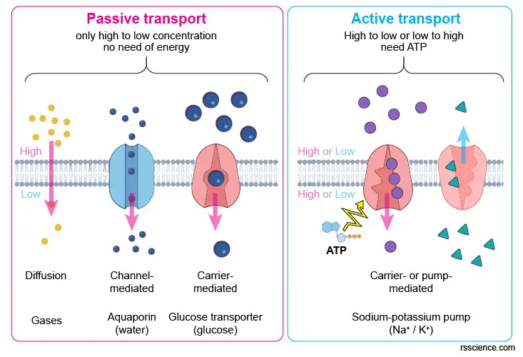
[In this figure] Depending on the nature of molecules, cells send them in either passive or active ways. Passive ship (diffusion or protein-mediated facilitated diffusion) only moves molecules from a high concentration to a lower one. Active transport requires special transporter proteins and uses free energy. Active transport tin can move substances in both directions.
ii. Active transport: Lipid bilayer finer repels many big, water-soluble molecules, and electrically charged ions, then cells must import or consign them to survive. Transport of these vital substances is carried out by certain classes of transmembrane proteins that function equally "pumps," which force solutes through the membrane when they are non full-bodied enough to diffuse spontaneously. In order to transport these substances, cells demand to spend energy or ATP. Adenosine triphosphate (ATP) is the biochemical energy "currency" of the cells.
Cells swallow and excrete by changing the jail cell membrane
Particles too large to be diffused or pumped are oftentimes swallowed or disgorged whole by an opening and closing of the membrane. To bring materials inside the cells, the cell membrane surrounding the particles undergoes invagination and engulfs past the formation of a vesicle (endocytosis). On the other hand, to remove unwanted materials from the cells, the membrane of a vesicle fuses with the cell membrane, extruding its contents to the surrounding medium, a process called exocytosis.

[In this figure] Endocytosis is the process in which cells absorb molecules by engulfing them. Exocytosis is a form of active ship in which a cell transports molecules out of the cell by secreting through the fusion of vesicle and cell membrane.
Cells talk to each other via direct or indirect contacts on their cell membranes
In a multicellular organism, the cells accept to communicate with each other. Cell-cell advice requires special proteins on the cell membranes. The jail cell that wants to ship a message has "ligand" proteins secreted or expressed on its cell membrane. And so, the recipient cells have respective "receptor" proteins to receive these letters (by bounden to the ligand proteins).
The recipient cells may respond immediately simply temporarily past irresolute prison cell shape or releasing certain ions. Alternatively, they may brand a slow but lasting change by passing the messages into the nucleus to plow on or plow off certain genes. When ii cells are close plenty, they may besides institute a directly substitution of molecules by protein channels (chosen gap junctions) spin the cell membranes of both cells.

[In this figure] Jail cell-jail cell communications via (a) straight contact and (b) gap junctions.
Photograph source: Tophat
Signal transduction along the cell membrane of nervous cells
Because the membrane acts equally a barrier for charged molecules and ions, they tin can occur in different concentrations on the two sides of the membrane. The deviation in full charge between the inside and outside of the jail cell is chosen the membrane potential.
For the nervous arrangement to function, neurons (or nervous cells) must be able to send and receive signals. These signals are electric currents generated by changing the membrane potential. The membrane potential tin alter in response to neurotransmitter molecules released from other neurons and ecology stimuli.
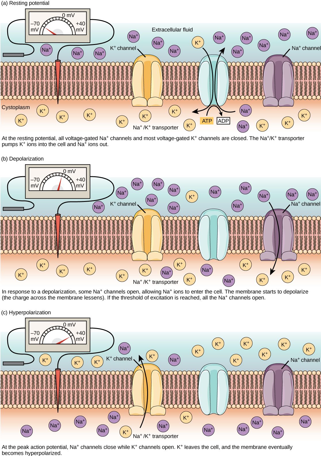
[In this figure] Voltage-gated ion channels open in response to changes in membrane potential.
The (a) resting membrane potential is a result of different concentrations of Na+ and Thousand+ ions within and outside the cell. A nerve impulse causes Na+ to enter the cell, resulting in (b) depolarization. At the tiptop activity potential, K+ channels open and the prison cell becomes (c) hyperpolarized.
Photo source: Lumenlearning
What does cell membrane look like under a microscope?
Under a compound light microscope, the cell membrane (only 5-10 nm) may be too sparse to exist seen. However, you can hands tell the boundary of cells if stained with proper dyes. That is where the cell membrane is.
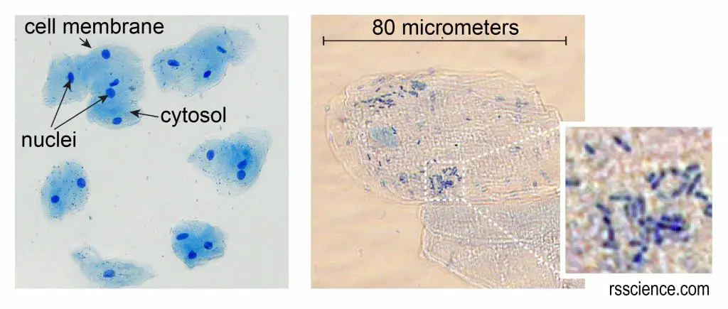
[In this effigy]Cheek cells stained with Methylene Blue.
The left image is a low magnification. You can run into the nuclei stained with a night bluish (because Methylene Blue stains DNA strongly). The cell membrane acts like a balloon and holds all the parts of a cell inside, such as a nucleus, cytosol, and organelles.
Plant cells have a layer of cell membrane underneath the cell wall. Most of the time, information technology is difficult to run across the jail cell membrane. However, you can find the prison cell membrane detached from the jail cell wall nether a hypertonic condition.

The cell membrane tin be stained with fluorescent dyes that demark to lipid. This provides a useful tool to visualize cell boundaries and morphology in multi-color staining experiments.
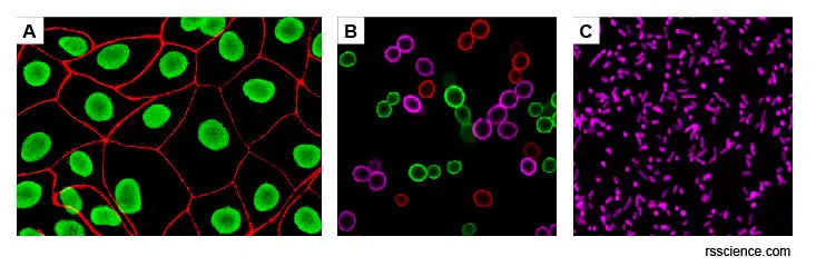
[In this figure] (A) Human epithelial cells stained for cell membrane (red) and nuclei (green); (B) Bakery'due south yeast cells (S. cerevisiae) stained with membrane dyes of three colors (ruby-red, royal, and green); (C) Bacteria, East. coli stained with royal membrane dyes.
Photo source: ABP Biosciences
Electron microscopes provide high-resolution images of cell membranes. Many important scientific findings were washed with electron microscopy.
Here are some examples:
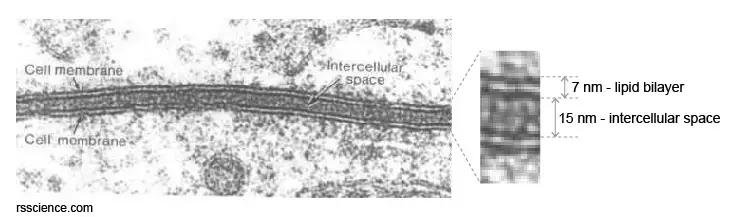
[In this effigy] Transmission electron microscopy (TEM) image of jail cell membranes of two very shut cells.
Photo source: cytochemistry.
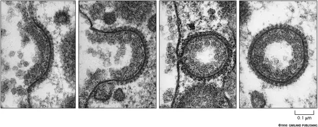
[In this figure] TEM paradigm of coated vesicle formation in endocytosis.
Photograph source: An Introduction to biological membranes
In order to study the structure of cell membrane, scientists used a "Freeze fracture" technique to tear apart the lipid bilayers and analyzed them separately nether a scanning electron microscope (SEM).
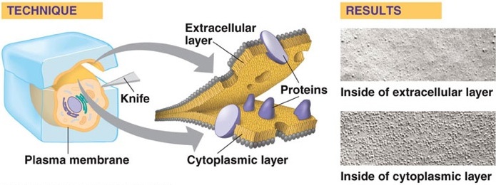
[In this figure] Freeze Fracture is a technique that tin can be used to visualize membrane construction and protein distribution.
First, a cell is rapidly frozen. Then it is cleaved along the fracture plane that splits the lipid bilayer. Separation forth this aeroplane exposes the transmembrane proteins embedded in the membrane. Right: the SEM micrographs show bumps on the surface of the split bilayer, which actually are transmembrane proteins. This confirmed that membrane proteins are randomly dispersed throughout the phospholipid bilayer. As well, in that location are integral transmembrane proteins that span the entire membrane.
Photo source: wikibooks
Special jail cell membrane structures in special cell types
To perform certain cellular functions, some cells possess unique jail cell membrane structures. Hither are some examples:
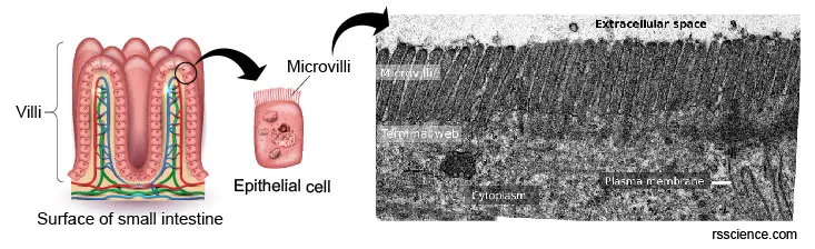
[In this figure] TEM image of the small intestine epithelium surface. Microvilli are microscopic cellular membrane protrusions that increment the surface area for maximizing nutrient absorption.
Photo source: Atlas of plant and animal histology.
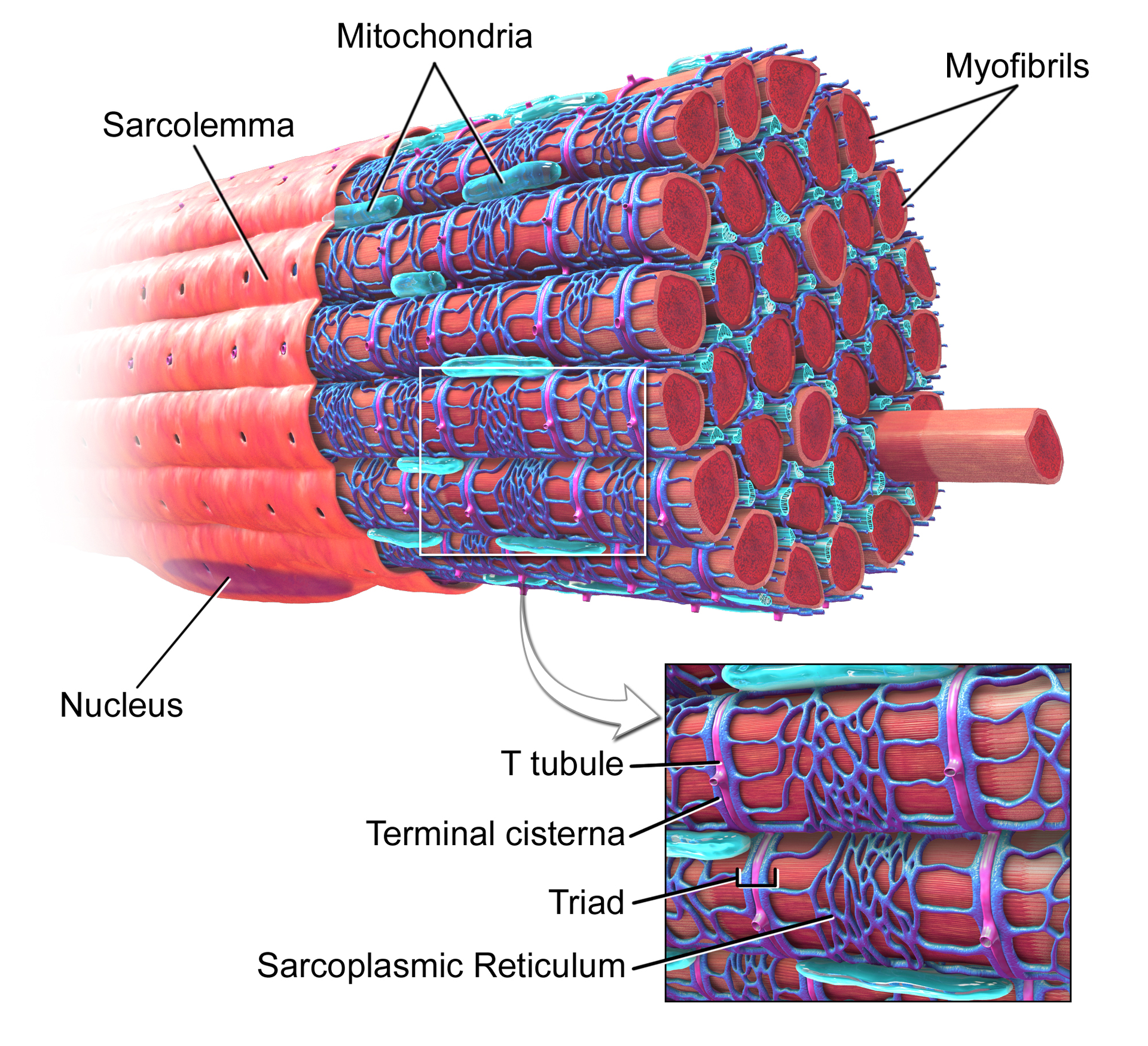
[In this figure] T-tubules (transverse tubules) are extensions of the cell membrane that penetrate into the center of skeletal and cardiac muscle cells. T-tubules permit the rapid manual of the activeness potential into the jail cell, allowing center muscle cells to contract more than forcefully.
Photo source: wiki
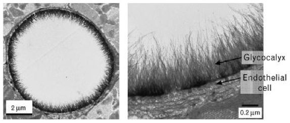
[In this figure] Electron micrograph showing the surface of endothelial cells' membrane coated with a thick layer of carbohydrate components, called glycocalyx. Endothelial cells are the prison cell types that line the inner lumens of blood vessels.
Photograph source: derangedphysiology
Summary
- Cell membrane is a biological membrane that separates the interior of the cell from the exterior space and protects the cell from its environment.
- Cell membrane is made past ii layers of lipid films (oil molecules) with many kinds of membrane proteins.
- Cell membrane controls the movement of molecules such as water, ions, nutrients, and oxygen in and out of the jail cell.
- Proteins on the cell membrane also involved in cell motion and cell-cell communication. For instance, cells received signals from outside through unlike kinds of receptor proteins on the cell membrane, performance like tiny antennas.
References
Source: https://rsscience.com/cell-membrane/
Posted by: hernandezflery1974.blogspot.com
0 Response to "What Is The Function Of The Cytosol In An Animal Cell"
Post a Comment